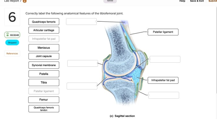
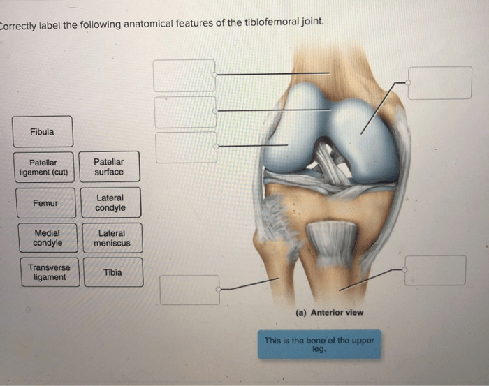
Correctly label the following anatomical features of the tibiofemoral joint. – Correctly labeling the anatomical features of the tibiofemoral joint is essential for understanding its function and pathology. This joint, formed by the articulation of the tibia and femur, is crucial for weight-bearing, movement, and stability of the knee.
This guide provides a comprehensive overview of the key anatomical structures of the tibiofemoral joint, including the tibial plateau, femoral condyles, menisci, cruciate ligaments, collateral ligaments, patella, and synovial membrane. By understanding the location, shape, and function of these components, we can gain a deeper appreciation of the complexity and resilience of the human knee joint.
Tibiofemoral Joint Anatomy

The tibiofemoral joint is a complex and essential articulation that connects the femur (thigh bone) to the tibia (shin bone). It is responsible for weight-bearing, stability, and mobility of the lower limb.
Tibial Plateau
The tibial plateau is the upper surface of the tibia. It consists of two concavities, the medial and lateral tibial plateaus, which articulate with the corresponding femoral condyles to form the tibiofemoral joint.
- Medial tibial plateau:Larger and more concave than the lateral plateau, it bears more weight during weight-bearing.
- Lateral tibial plateau:Smaller and less concave than the medial plateau, it provides stability to the joint.
Femoral Condyles, Correctly label the following anatomical features of the tibiofemoral joint.
The femoral condyles are the rounded projections at the distal end of the femur. They are covered with articular cartilage and articulate with the tibial plateaus to form the tibiofemoral joint.
- Medial femoral condyle:Larger and more concave than the lateral condyle, it articulates with the medial tibial plateau.
- Lateral femoral condyle:Smaller and less concave than the medial condyle, it articulates with the lateral tibial plateau.
Menisci
The menisci are two C-shaped fibrocartilaginous structures located between the tibial plateaus and the femoral condyles.
- Medial meniscus:Larger and more crescent-shaped than the lateral meniscus, it attaches to the medial collateral ligament.
- Lateral meniscus:Smaller and more circular than the medial meniscus, it attaches to the popliteus muscle.
The menisci provide shock absorption, joint stability, and distribute load evenly across the joint surface.
Cruciate Ligaments
The cruciate ligaments are two strong bands of connective tissue that cross each other within the tibiofemoral joint.
- Anterior cruciate ligament (ACL):Prevents excessive anterior translation of the tibia relative to the femur.
- Posterior cruciate ligament (PCL):Prevents excessive posterior translation of the tibia relative to the femur.
Collateral Ligaments
The collateral ligaments are two bands of connective tissue located on either side of the tibiofemoral joint.
- Medial collateral ligament (MCL):Prevents excessive varus (inward) angulation of the knee joint.
- Lateral collateral ligament (LCL):Prevents excessive valgus (outward) angulation of the knee joint.
Patella
The patella, also known as the kneecap, is a small, triangular bone that sits anteriorly to the tibiofemoral joint.
It articulates with the trochlear groove of the femur, which allows for smooth knee extension and flexion.
Synovial Membrane
The synovial membrane is a thin layer of tissue that lines the inner surface of the tibiofemoral joint.
It secretes synovial fluid, which lubricates the joint and reduces friction during movement.
FAQs: Correctly Label The Following Anatomical Features Of The Tibiofemoral Joint.
What is the function of the tibial plateau?
The tibial plateau provides a stable and congruent surface for articulation with the femoral condyles, facilitating weight-bearing and joint stability.
How do the cruciate ligaments prevent excessive movement of the tibia?
The anterior cruciate ligament (ACL) prevents excessive anterior translation of the tibia, while the posterior cruciate ligament (PCL) prevents excessive posterior translation.
What is the role of the menisci in the tibiofemoral joint?
The menisci are fibrocartilaginous structures that provide shock absorption, joint stability, and nutrient distribution within the joint.


